Pierre Bon
Scalar Electromagnetic Field imaging without reference and with nanoscale resolution on living biological samples

Date & heure
06/11/2024 – 11 am
Lieu
4 place Jussieu 75005
Amphi Astier
Accueil
A coffee will be offered starting at 10:45 am, the seminar will start at 11am
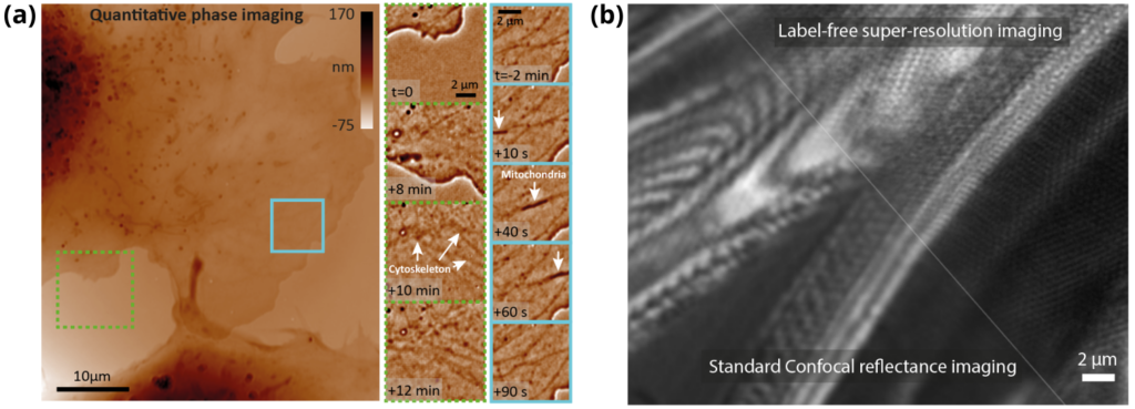
Accessing the phase of a light beam in addition to its intensity drastically increases the amount of information that can be extracted from light. The development of compact and self-referenced interferometry solutions1 has opened the field to high-resolution measurement of the scalar electromagnetic field on any type of beam without the need for an external reference. This modality proves to be particularly interesting as it allows measurement of optical path differences as small as 10pm (≈λ/50 000) in single shot. Applied to microscopy2 of semi-transparent biological samples, it allows to infer biophysical information including their mass3, nature4, position5,6, or even the presence of temperature variations7 at very high resolution.
Recently, to push the resolution of quantitative phase imaging beyond limit, we have proposed a solution to bypass the diffraction limit (so-called super-resolution imaging) on any sample8,9. This approach now enables the imaging of electromagnetic fields with a resolution better than 100 nm while remaining compatible with the observation of fragile 3D samples such as organoids.
1. Primot, J. et al. Achromatic three-wave (or more) lateral shearing interferometer. J Opt Soc Am A 12, 2679 (1995).
2. Bon, P. et al. Quadriwave lateral shearing interferometry for quantitative phase microscopy of living cells. Opt Express 17, 13080–13094 (2009).
3. Aknoun, S. et al. Living cell dry mass measurement using quantitative phase imaging with quadriwave lateral shearing interferometry: an accuracy and sensitivity discussion. J. Biomed. Opt. 20, 126009 (2015).
4. Nguyen, M.-C. et al. Label-Free Single Nanoparticle Identification and Characterization in Demanding Environment, Including Infectious Emergent Virus. Small n/a, 2304564 (2023).
5. Bon, P. et al. Self-interference 3D super-resolution microscopy for deep tissue investigations. Nat. Methods 15, 449–454 (2018).
6.Bon, P. et al. Three-dimensional nanometre localization of nanoparticles to enhance super-resolution microscopy. Nat Commun 6, 7764- (2015).
7. Baffou, G. et al. Thermal Imaging of Nanostructures by Quantitative Optical Phase Analysis. ACS Nano 6, 2452–2458 (2012).